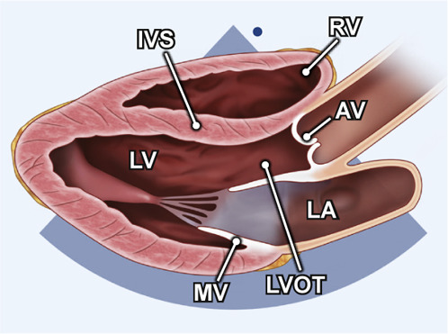
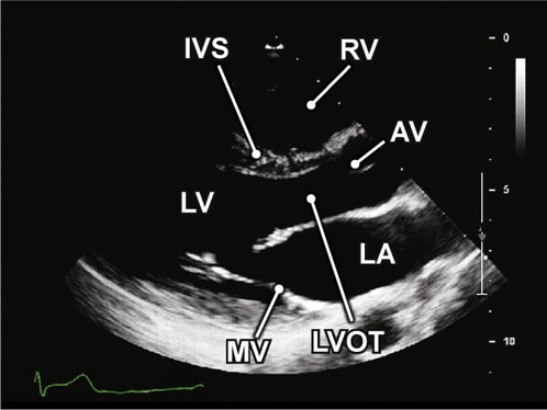
Reduce depth to display only LV, LA, MV. AOV. and RV
Acquisition: Parasternal window PLAX view Left sternal border, transducer orientation toward right shoulder, beam positioned perpendicular to left ventricle
Structures to demonstrate: LA MV LV LVOT AV IVS RV
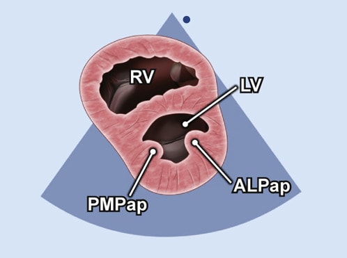
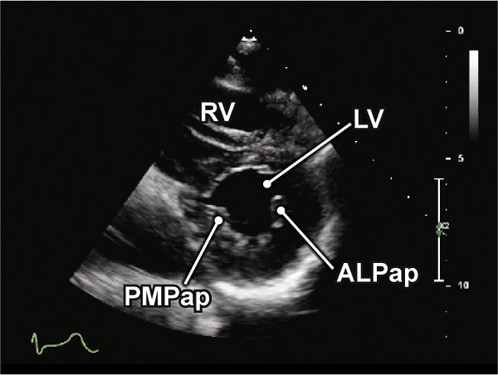
Parasternal Short Axis
2D loop of LV at mid papillary level
Acquisition: Parasternal window PSAX view. Tilt inferiorly from MV
Structures to demonstrate: RV IVS PMPap ALPap LV
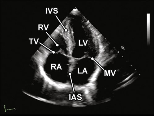
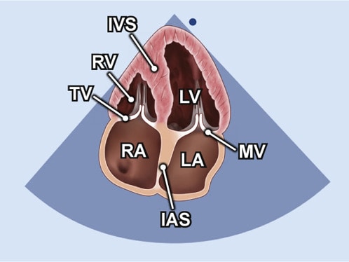
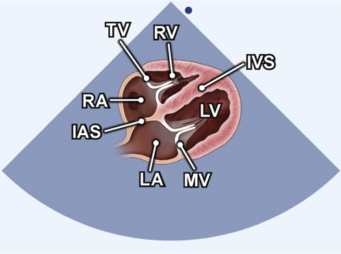
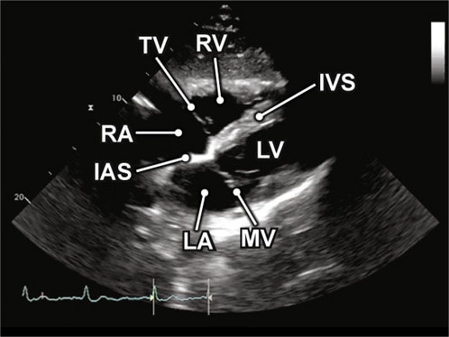
Apical 4 Chamber
2D view at at optimal depth to display all 4 cardiac chambers
Acquisition: Move to patient’s left side to identify apical impulse, align orientation toward bed
Structures to demonstrate: LA MV LV RA RV TV IVS IAS
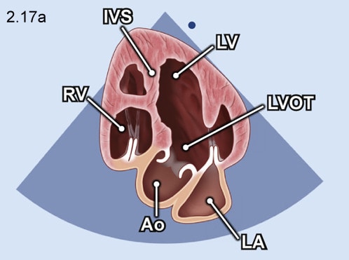
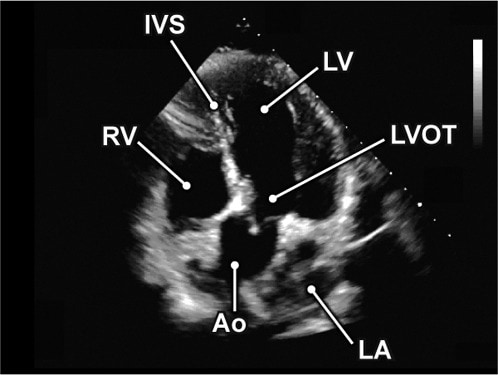
Apical 5 Chamber
2D view to show all five chambers
Acquisition: From the A4C view tilt the beam anteriorly to show the LVOT
Structures to demonstrate: LA MV LV IVS LVOT RA RV
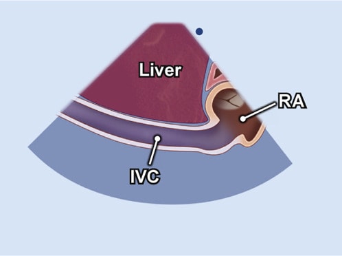
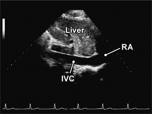
Subcostal 4 Chamber
2D view of all 4 chambers
Acquisition: Patient supine with transducer at subxiphoid position, orientation marker toward patient’s left shoulder
Structures to demonstrate: LA MV LV LVOT AV IVS RV
Prepared for CardioGuide by Atul Jaidka. Images and videos from ASE 2019 Guidelines for Performing a Comprehensive Transthoracic Echocardiographic Examination in Adults: Recommendations from the American Society of Echocardiography