Introduction
- Procedure to remove fluid from the pericardial space for diagnostic and therapeutic purposes.
- This is considered a lifesaving procedure for patients presenting with cardiac tamponade.
Techniques to access pericardial space
- Blind Procedure: Rarely performed with the advent of ultrasound. Can be done in a code blue or trauma situation. This procedure is typically done via subcostal approach.
- Static Ultrasound: Using the ultrasound, visualize the best fluid pocket and estimate the angle of approach for needle insertion. Needle is inserted without direct ultrasound guidance.
- Dynamic Ultrasound: Direct visualization of the needle entering the pericardium. This technique is technically challenging due to the need to visualize the needle in-plane with the ultrasound beam and guide it into the pericardial space. (this is the technique we teach on CardioGuide.ca!)
- Fluroscopically guided: Usually done in the cath lab with direct visualization of the needle with x-ray guidance. Radio-contrast is injected through the needle to confirm position in the pericardial space.
Dynamic Ultrasound Technique Video
NOTE: Recommend watching the video in full-screen mode
Steps
- Confirm window:
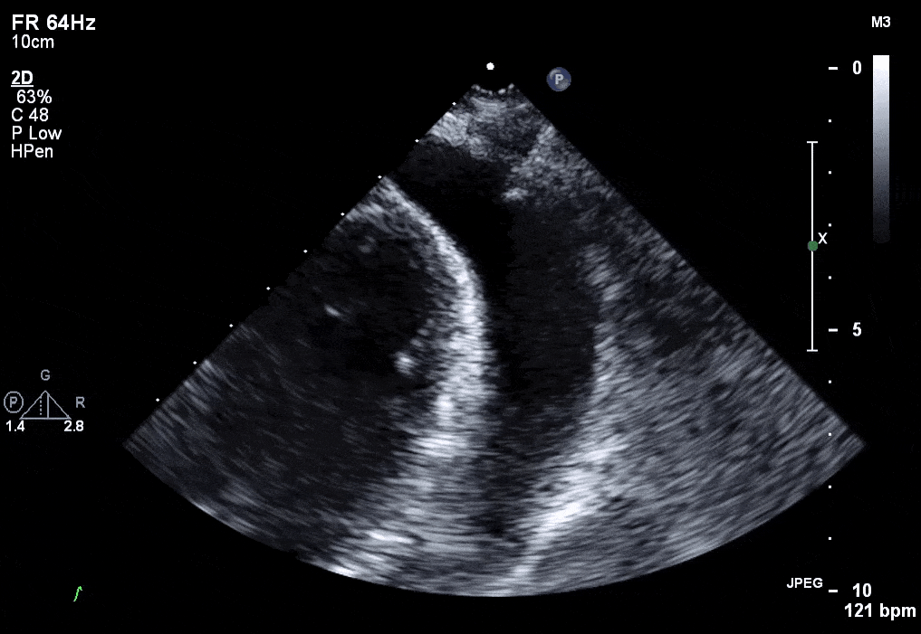
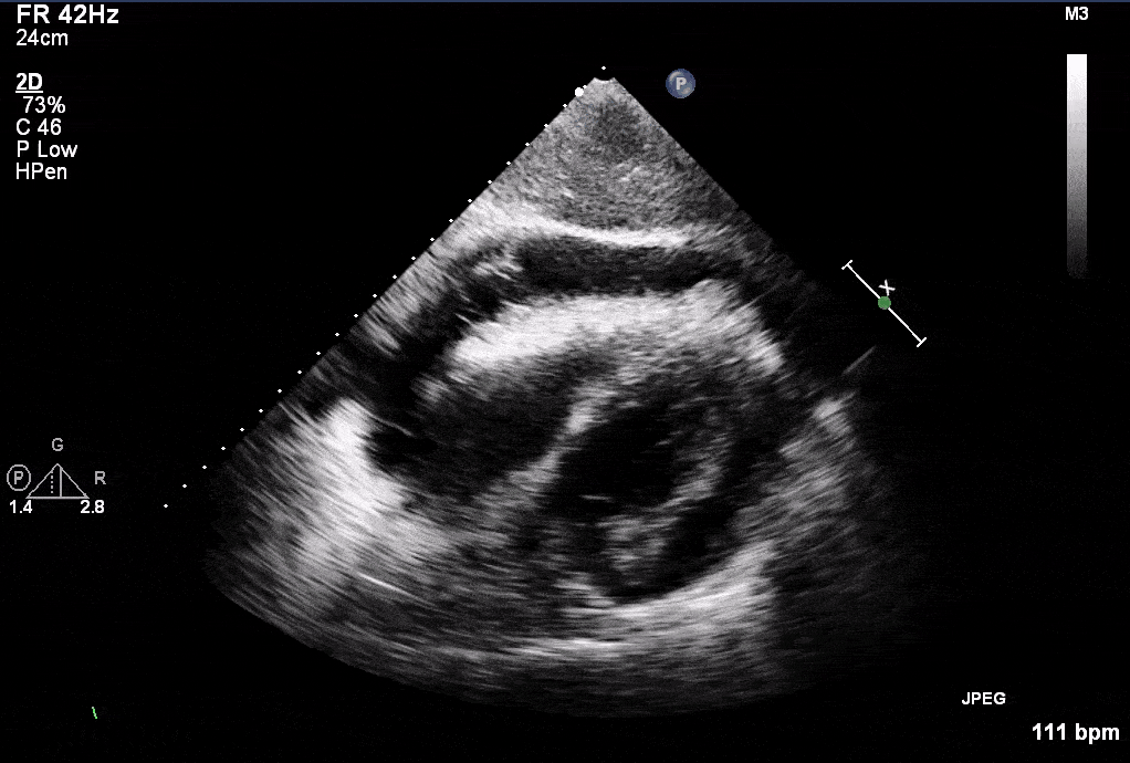
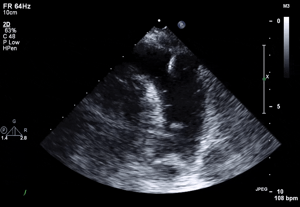
- Ultrasound patient to select the appropriate window (apical, parasternal, or subcostal), and identify relevant structures.
- Practical tip: Use the window that allows access to the largest pocket of fluid that is clear of other structures to reduce risk of complications (ie. hepatic laceration or pneumothorax)
- Preparation:
- Collect supplies and position the patient supine with the head of the bed raised to 30-45 degrees
- Sterilize the skin and create a sterile environment
- Anesthetize Skin
- Freeze skin with 1-2% lidocaine to anesthetize the skin overlying the needle entry site.
- Micropuncture needle insertion
- The micropuncture needle is inserted under dynamic in-plane ultrasound guidance. Periodic or continuous negative pressure is applied to detect when the needle tip enters the pericardial space.
- Practical tip: Use syringe with lidocaine and inject to anesthetize deeper structures while advancing the needle under ultrasound visualization
- Exchange for sheath
- Using seldinger technique, exchange needle for sheath with corresponding wire (included in the micropuncture kit)
- Practical tip: If serosanguinous or bloody effusion, a sample can be left in a plastic tray to check for clotting.
- Bubble study
- Using 3-way stopcock attached to sheath, infuse agitated saline, and visualize with ultrasound in the same or alternate window.
- Drainage Catheter insertion
- Using seldinger technique again, exchange sheath for catheter using J tip wire
- Practical tip: skin nick and catheter dilator helps facilitate catheter insertion
- Drain fluid
- Practical tip: Use syringe to drain fluid initially to achieve hemodynamic stability, send for samples and then attach to drainage system
Confirmation Tests
- Very important to confirm placement prior to dilation and insertion of drain.
- Dilation and insertion of the drainage catheter into the ventricle is a catastrophic complication.
- How to confirm: (recommended to use multiple methods)
- Direct visualization of the needle entering the pericardial space
- Visualizing the guidewire in the pericardial space
- Agitated Saline Injection (recommended)
- Saline is agitated using two syringes connected with a 3-way stopcock. The agitated saline is is injected quickly into the needle or micropuncture catheter, while imaging the pericardial space with ultrasound.
- Clotting Test
- Place a small amount of pericardial fluid in a plastic container (often equipment tray). Most pericardial fluid should not form clots and should remain liquid.
- Authors: Dr. Atul Jaidka, Dr. Daniel Durocher and Dr. Pavel Antiperovitch (MD, FRCPC, Cardiology Fellow)